

")
")
")
")

")
")
")
")


Description
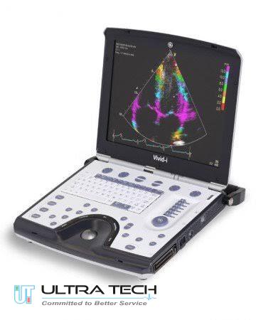
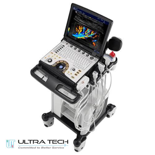
GE NextGen Logiq e Ultrasound Machine
GE NextGen LOGIQ E Ultrasound Machine(for ICU,NICU,CCU,OT)
Overview
The GE NextGen Logiq e is a compact and powerful portable ultrasound machine designed for Point-of-Care (POCUS), musculoskeletal (MSK), vascular, anesthesia, emergency medicine, and general imaging applications. The system delivers console-level imaging performance in a lightweight laptop-style design, making it ideal for hospitals, clinics, operating rooms, and mobile healthcare environments.
The latest LOGIQ e platform offers advanced imaging technologies, AI-assisted workflow tools, strain elastography, and enhanced portability for fast and confident diagnosis.
Key Components & Concepts
Transducer (Probe): The part that touches the skin, emits sound, and receives echoes; different probes are used for different body parts/depths.
Gain: Controls the brightness/contrast of the image (adjusts overall image sensitivity).
Focus: Narrows the sound beam at a specific depth to improve detail.
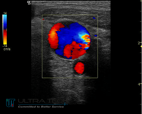
Modes: Different displays like B-mode (brightness/2D), M-mode (motion), and Doppler (blood flow color-coded).
Why It’s Used (Applications)
Obstetrics/Gynecology: Pregnancy scans, ovarian cysts, fibroids.
Cardiology: Heart function, blood flow.
Vascular: Detecting clots, narrowing/widening of blood vessels.
Abdominal: Examining liver, kidneys, and gallbladder.
Procedural Guidance: Guiding needles for biopsies or anesthesia.
Safety
Generally considered safe and non-invasive.
Key Features of GE NextGen Logiq e
- Portable laptop-style ultrasound system
- High-quality 2D and Color Doppler imaging
- CrossXBeam™ spatial compound imaging
- SRI-HD (Speckle Reduction Imaging)
- AI Needle Recognition technology
- Strain Elastography support
- Scan Assistant workflow automation
- Tissue Harmonic Imaging (THI)
- B-Mode, M-Mode, Color Flow Imaging
- Power Doppler and Spectral Doppler
- Lightweight and mobile design
- Up to 90-minute battery operation
- 15.6-inch high-resolution monitor
- Sealed keyboard for easy cleaning
- Trackball and trackpad control
- DICOM connectivity support
- Fast boot-up and workflow optimization
- Specialized POCUS and intervention tools
- ATO [Automatic Tissue Optimization]
- Raw data processing
- Anatomical M-Mode
- PW Doppler
- CW Doppler,
- Tissue Harmonics
- On-board patient data management
- ComfortScan
- SmartScan
- TruAccess
- Advanced Image compare
- Image archiving
- CINE loop
- 80GB HD
- CD-RW
- 2 USB ports
- Wireless LAN image transfer
GE NextGen Logiq e Ultrasound Options:
- DICOM
- Speckle Reduction Imaging [SRI]
- CrossXBeam
- Freehand 3D
- CW Doppler
- LogiqView
- ECG Module
Transducer types of GE NextGen Logiq e |
||
| Convex array
|
C1-5-RS, 4C-RS | Abdominal, fetal/obstetrics, urology (including prostate), pediatric, neonatal cephalic, nerve block, musculoskeletal conventional/superficial, thoracic/pleural, tissue biopsy |
| Microconvex array | 8C-RS, E8C-RS
|
Abdominal, cardiac (adult and pediatric), peripheral vascular, pediatric, neonatal cephalic, small organ, nerve block, musculoskeletal conventional/ superfical, ophthalmic, thoracic/pleural |
| Linear array | ML6-15-RS, 9L-RS, 12L-RS, L4-12t-RS, L8-18i-RS, L10-22-RS, L4-20t-RS
|
Peripheral vascular, pediatric, small organ, neonatal cephalic, nerve block, musculoskeletal conventional/superficial, thoracic/pleural, ophthalmic, vascular access (IV, PICC), tissue biopsy |
| Phased array | 3Sc-RS, 6S-RS, 12S-RS, M5SC-RS1
|
Abdominal, fetal/obstetrics, cardiac (adult and pediatric), peripheral vascular, urology (including prostate), pediatric, neonatal cephalic, adult cephalic, thoracic/pleural, ophthalmic, tissue biopsy |
| TEE | 6Tc-RS | Cardiac |
Clinical Applications of GE NextGen Logiq e
Point of Care Ultrasound (POCUS)
- ICU
- NICU
- CCU
- COT
- Critical Care
- Emergency Medicine
- Bedside Ultrasound
Anesthesia
- Nerve Blocks
- Regional Anesthesia
- Pain Management
Musculoskeletal (MSK)
- Tendon Assessment
- Sports Medicine
- Joint Imaging
- Rheumatology
Vascular Imaging
- DVT Assessment
- Carotid Studies
- Peripheral Vascular Imaging
General Imaging
- Abdomen
- Small Parts
- Thyroid
- Urology
- Soft Tissue Imaging
Advanced Imaging Technologies
CrossXBeam™ Imaging
Provides enhanced tissue border definition and improved visualization through spatial compounding technology.
SRI-HD Technology
Reduces image noise and speckle artifacts for improved tissue visualization and diagnostic confidence.
AI Needle Recognition
Helps clinicians visualize needle position during interventional procedures and regional anesthesia.
Strain Elastography
Supports tissue characterization and treatment monitoring by evaluating tissue stiffness.
Specifications of GE NextGen Logiq e
| Feature | Details |
| System Type | Portable Laptop Ultrasound |
| Display | 15.6″ High-Resolution LCD |
| Imaging Modes | B, M, Color Doppler, Power Doppler, PW Doppler |
| Applications | POCUS, MSK, Vascular, Anesthesia, General Imaging |
| Battery Life | Up to 90 Minutes |
| Connectivity | DICOM, USB, Network |
| Weight | Approx. 5.16 kg |
| Probe Support | Linear, Convex, Cardiac, Specialty Probes |
| Workflow Tools | Scan Assistant, AI Needle Recognition |
Application Probes
Linear Probes
- L4-12t-RS
- L8-18i-RS
- L4-20t-RS
Convex Probes
- C1-5-RS
Cardiac Probes
- 12S-RS
- 3Sc-RS
Why Choose GE NextGen Logiq e?
Excellent Portability
Compact laptop design allows easy transport between departments and bedside examinations.
Advanced Imaging Quality
Delivers high-resolution ultrasound imaging with advanced image enhancement technologies.
Fast Workflow
Scan Assistant and AI-powered tools help reduce examination time and improve efficiency.
Ideal for Interventional Procedures
Needle recognition and MSK imaging capabilities make it suitable for anesthesia and procedural guidance.
Why Buy from Ultratech BD ?
- Professionally tested ultrasound systems
- Installation and application training
- Technical service support
- Genuine probes and accessories
- Competitive pricing in Bangladesh
- Experienced medical equipment team
Warranty & Support in Bangladesh
At Ultratech BD refurbished GE Voluson S8 ultrasound systems may include:
- Installation support
- Application training
- Technical servicing assistance
- Preventive maintenance support
- Probe support and repair assistance
- After-sales engineering support
- Online and onsite troubleshooting support
- Warranty coverage depending on machine condition and package configuration
For detailed warranty duration and support availability in Bangladesh, contact directly before purchase.
Frequently Asked Questions (FAQ) – GE NextGen Logiq e ?
What is the GE NextGen Logiq e used for?
The GE LOGIQ e is used for POCUS, musculoskeletal imaging, vascular studies, anesthesia guidance, emergency medicine, and general ultrasound applications.
Does GE LOGIQ e support Doppler imaging?
Yes, it supports Color Doppler, Power Doppler, and Spectral Doppler imaging.
Is the system portable?
Yes, it is a lightweight laptop-style ultrasound system with battery-powered operation.
Does it support needle guidance?
Yes, the system includes AI Needle Recognition technology for interventional procedures.
What is the battery backup time?
The battery can operate for approximately 90 minutes between charges.
What does Ultratech BD provide?
Ultratech BD supplies high-quality new and refurbished medical equipment including ultrasound machines, endoscopy systems, patient monitors, OT equipment, ECG machines, X-ray systems, CT scanners, MRI machines, and laboratory equipment.
Does Ultratech BD sell refurbished medical equipment?
Yes. Ultratech BD offers professionally tested and refurbished medical equipment from leading international brands such as GE Healthcare, Olympus, Karl Storz, Stryker, Hitachi, and others.
Which ultrasound machine brands are available?
Ultratech BD provides ultrasound systems from GE Healthcare including LOGIQ and Voluson series models.
Do you provide installation services?
Yes. Installation, system setup, and basic operational guidance are available for medical equipment purchased from Ultratech BD.
Is technical support available after purchase?
Yes. Ultratech BD offers technical support and service assistance for various medical devices and diagnostic systems.
Do you provide warranty support?
Warranty availability depends on the product type and condition. Customers can contact Ultratech BD for specific warranty information before purchase.
Where is Ultratech BD located?
Ultratech BD is located at:
1282, East Monipur, Begum Rokeya Sarani Road, Mirpur, Opposite of Al Helal Hospital, Near Metro Pillar No-266, Dhaka-1216, Bangladesh.
How can I contact Ultratech BD?
You can contact Ultratech BD through:
- Engineer Support: +880 1717 670 039
- Office: +880 170 7078 538
- Email: info@ultratechbd.com
Does Ultratech BD supply endoscopy equipment?
Yes. Ultratech BD offers Olympus endoscopy systems including CV-170, CV-150, CV-260/CLV-260, and related accessories.
Do you provide OT and surgical equipment?
Yes. Ultratech BD supplies OT lights, OT tables, insufflators, electrosurgical units, laparoscopy systems, and surgical accessories.
Are your products suitable for hospitals and diagnostic centers?
Yes. The equipment is suitable for hospitals, clinics, diagnostic centers, imaging facilities, and specialized medical practices.
Can I request a quotation for medical equipment?
Yes. Customers can contact Ultratech BD directly for product availability, pricing, and customized quotations.
Does Ultratech BD offer nationwide delivery in Bangladesh?
Product delivery and logistics support may be available depending on equipment type and location. Contact the sales team for delivery details.
How long has Ultratech BD been operating?
Ultratech BD was established in Bangladesh in 2002 and has experience in supplying branded medical equipment and healthcare technology solutions.
Official Website


admin –
VERY Good